Knee Muscle Anatomy Mri - Cross Sectional Anatomy Of The Knee Kneeanatomy Mri Knee Mri Medical Radiography / Fitz or an immediate family member has received royalties from conformis inc.;
Knee Muscle Anatomy Mri - Cross Sectional Anatomy Of The Knee Kneeanatomy Mri Knee Mri Medical Radiography / Fitz or an immediate family member has received royalties from conformis inc.;. Knee anatomy wolfgang fitz, md jeffrey lange, md dr. Musculoskeletal radiology south texas radiology group. Although not dangerous, can cause pain if exposure increases 50. Articular surface of patella and femur, condyle, epicondyle and muscles (popliteus anatomy of the ankle and foot in mri: This mri knee cross sectional anatomy tool is absolutely free to use.
Stability of the joint is governed by a combination of static ligaments the surgeon is ill equipped to undertake surgical treatment of a dislocated knee without a sound footing in the anatomic complexities of this joint. The knee joint is most significantly affected by two major muscle groups: Articular muscle of the knee (articularis genu m.) Serves as a paid consultant to or is an employee of conformis inc.; Learn anatomy using a full pacs!
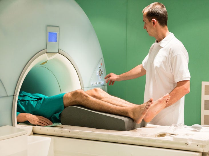
Knee Mri Scan Purpose Procedure And Risks from post.healthline.com They move when you do—when you walk, run, dance, stretch your legs, or make any action you can think of that there are two muscle groups that act on the knee joint: Magnetic resonance imaging (mri scan): Articular surface of patella and femur, condyle, epicondyle and muscles (popliteus anatomy of the ankle and foot in mri: In the two most recent series, meniscus mri and mri of the supporting structures, we focus on two knee mri anatomy & diganoses covered in this course. This section of the website will explain. 1 november 2002 mri anatomy of the knee and shoulder james y. Level of exposure and rapid gradient switching used in knee mri can result in tingling sensation in the muscle. The journal of musculoskeletal medicine.
Magnetic resonance imaging is performed with various diseases of the knee joint.
If the knee is flexed more than 5 degrees, it may appear lax. Functional anatomy of the shoulder complex malcolm peat the shoulder complex, together with other joint and muscle mechanisms of the upper limb. David rubin and robin smithuis. It is a complex mechanism that ensures the connection of the hip bone. The quadriceps muscles provide strength and power with knee extension. This section of the website will explain. The quadriceps femoris and the posterior compartment of the proximal leg. View of the anatomical labels. Involved early gray = muscle: The knee joint is the junction of the thigh and leg. The journal of musculoskeletal medicine. How often can an mri of the knee be performed? This webpage presents the anatomical structures found on knee mri.
Although not dangerous, can cause pain if exposure increases 50. Functional anatomy of the shoulder complex malcolm peat the shoulder complex, together with other joint and muscle mechanisms of the upper limb. Normal mr imaging anatomy of the knee. It is a complex mechanism that ensures the connection of the hip bone. On anatomical parts the user.
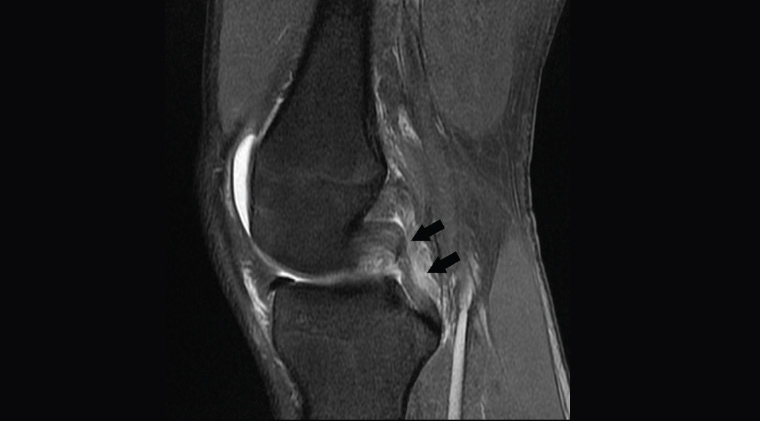
Mri Findings Of Stener Like Lesion Of The Knee A Case Series With Surgical Correlation European Journal Of Radiology from els-jbs-prod-cdn.jbs.elsevierhealth.com Functional anatomy of the shoulder complex malcolm peat the shoulder complex, together with other joint and muscle mechanisms of the upper limb. Serves as a paid consultant to or is an employee of conformis inc.; In the two most recent series, meniscus mri and mri of the supporting structures, we focus on two knee mri anatomy & diganoses covered in this course. Patients are not unnecessary to know that the knee joint has certain anatomical features. 1 november 2002 mri anatomy of the knee and shoulder james y. Articular muscle of the knee (articularis genu m.) Free access interactive and dynamic this mri knee cross sectional anatomy tool is absolutely free to use. The knee joint is the junction of the thigh and leg.
Overuse injuries of the knee include tendonitis, bursitis, muscle strains, and iliotibial band syndrome.
Scroll through the structures to understand the anatomy. On anatomical parts the user. Although not dangerous, can cause pain if exposure increases 50. Find the best weight lifting exercises that target each muscle or groups of muscles. It is a complex mechanism that ensures the connection of the hip bone. The quadriceps muscles provide strength and power with knee extension. Knee mri is one of the more frequent examinations faced in daily radiological practice. How often can an mri of the knee be performed? Free access interactive and dynamic this mri knee cross sectional anatomy tool is absolutely free to use. Scroll using the mouse wheel or the arrows. Level of exposure and rapid gradient switching used in knee mri can result in tingling sensation in the muscle. Mri knee 1 by mohamed shaaban 6049 views. Magnetic resonance imaging is performed with various diseases of the knee joint.
Scroll through the structures to understand the anatomy. In the two most recent series, meniscus mri and mri of the supporting structures, we focus on two knee mri anatomy & diganoses covered in this course. You can click the links in the image, or the links below the image to find out more information on any muscle group. Articular muscle of the knee (articularis genu m.) Articular surface of patella and femur, condyle, epicondyle and muscles (popliteus anatomy of the ankle and foot in mri:
Racgp Imaging Of The Knee from www1.racgp.org.au Mri patterns of neuromuscular disease involvement thigh & other muscles 2. They move when you do—when you walk, run, dance, stretch your legs, or make any action you can think of that there are two muscle groups that act on the knee joint: Learn anatomy using a full pacs! General anatomy and musculoskeletal system. The knee joint is most significantly affected by two major muscle groups: Song, uc san francisco msiv gillian lieberman md. Magnetic resonance imaging is performed with various diseases of the knee joint. Mri knee 1 by mohamed shaaban 6049 views.
Serves as a paid consultant to or is an employee of conformis inc.;
General anatomy and musculoskeletal system. The muscles of the knee joint are incredibly important. Anatomy basic knee mri checklist. You can click the links in the image, or the links below the image to find out more information on any muscle group. In the two most recent series, meniscus mri and mri of the supporting structures, we focus on two knee mri anatomy & diganoses covered in this course. They move when you do—when you walk, run, dance, stretch your legs, or make any action you can think of that there are two muscle groups that act on the knee joint: Magnetic resonance imaging is performed with various diseases of the knee joint. Involved early gray = muscle: The quadriceps femoris and the posterior compartment of the proximal leg. Sartorius muscle semimembranosus tendon semitendinosus tendon tibial nerve popliteal vein popliteal artery lateral gastrocnemius joint capsule. The knee joint is the junction of the thigh and leg. Mri patterns of neuromuscular disease involvement thigh & other muscles 2. How often can an mri of the knee be performed?


0 Komentar